Intended for healthcare professionals
Open access
Research article
First published online May 19, 2023

Comparative Assessment of Success Rate Between Silver Nanoparticle Coated and Uncoated Infra-zygomatic Bone Screws: A Split-Mouth Study

Abstract

Introduction

In orthodontic treatment, infra-zygomatic crest screws have been effectively used in the correction of borderline skeletal malocclusion. The success of bone screws depends on factors such as biomechanical factors, placement technique, and microbial factors. Therefore, this study aimed to evaluate the success rate as well as assess the microbiota of silver nanoparticle coated and uncoated infra zygomatic crest (IZC) screws on clinical application.

Method

A sample of 36 stainless steel IZC screws was divided into 2 groups. Group I- Silver nanoparticle (AgNP) coated (n = 18) and group II uncoated (n = 18). A total of 18 patients (4 male, 14 female; mean age 22.58±3.52) received IZC screws (coated or uncoated) via a double-blind, split-mouth design. The success rate was evaluated based on the stability of IZC screws. And also molecular analysis was carried out using terminal restriction fragment length polymorphism complemented with cloning and identification of species.

Results

The success rate was higher in group I (77.80%) than in group II (50.00%). The mean rank for terminal restriction fragments in AgNP coated group (14.92%) was significantly lower than the uncoated group (22.08%) (P<0.05). The cloning results revealed a major difference in the predominant species composition between AgNP-coated (Prevotella oris sp.) and uncoated IZC screw (Capnocytophaga leadbetteri).

Conclusion

The success rate for AgNP-coated IZC screws was higher compared to uncoated screws, while predominant bacteria present in coated and uncoated samples were found to be of different biological significance which needs to be explored subsequently.

Introduction

The infra zygomatic crest (IZC) bone screws are effective skeletal anchorage devices used in orthodontics for en-mass retraction, intrusion, and distalization of the maxillary arch. It is also effective in treating borderline surgical cases to nonsurgical ones without compromising the results. However, IZC screws pose a few challenges, most common being uncontrollable soft tissue inflammation, gingival overgrowth on the screw, and early loosening of the screw.1
According to a study, 43% of individuals with implants suffer from peri-implantitis.2 Peri-implant inflammation caused by plaque retention leads to mobility and loss of the orthodontic bone screws. In a study, Sato et al.3 reported that an anaerobic environment in crevices around orthodontic anchor plates is favorable for the growth of anaerobes. The knowledge of quantitative and qualitative aspects of predominant microorganisms present in the peri-implant region is important for reducing inflammation.4 Reports suggest gram-negative obligate anaerobes are predominantly comprised of bacterial flora in peri-implantitis.5
In the recent past to reduce or nullify the adhesion of microbes onto the surface of implants many methods like surface treatments such as polishing, coating, and modification of surface free energy, have been employed. 6
In this regard, silver nanoparticles are of prime interest in surface coating procedures due to their high antimicrobial actions against both gram-positive and gram-negative bacteria, viruses, and other eukaryotic microorganisms compared to other metal nanoparticles.7 Evidence from previous in-vitro6, 8 and in-vivo studies911 on AgNP-coated titanium mini-implant, orthopedic implants as well as stainless steel (SS) orthodontic brackets, and orthodontic band cement have confirmed that AgNP gives antibacterial and anti-adhesive characteristics. However, there are no reported studies comparing success rate as well as microbiota between AgNP-coated and uncoated SS IZC bone screws on clinical application. SS is the material of choice for bone screws rather than titanium or titanium alloy because of its toughness when placed in the cortical bone.12 In the current literature the controversy remains regarding the effectiveness of in-vitro studies because of various limitations like the inability to reproduce oral environment, variation in bone density, and also various other hormonal and systemic conditions which will influence the outcome of the study.13
Therefore this study aimed to surface coat SS IZC screws with AgNP and evaluate the success rate as well as microbiota of silver nanoparticle coated and uncoated infra-zygomatic crest (IZC) screws on clinical application. The study proceeded with the assumption that there was no difference in success rate between AgNP-coated and uncoated IZC screws on clinical application.

Materials and Methods

The study was approved by the Institutional Ethical Committee (No:110/2017-18) to compare the success rate for AgNP-coated and uncoated IZC screws. This double-blind split-mouth study included 18 patients (4 male, 14 female; mean age 22.58 ± 3.52) undergoing fixed orthodontic treatment with MBT mechanotherapy (3M Unitek, 0.022″ slot) requiring bilateral IZC anchorage to correct Class II malocclusion. The patients were selected based on the following selection criteria, after obtaining informed consent:
Cases with congenitally missing third molars and/or samples where the third molars were removed before the treatment were considered for the study.
Patients with good oral hygiene and no evidence of periodontal bone loss after radiographic examination with post-alignment overjet of 4.8±0.6 (mm) and overbite of 5.8 ± 0.9 (mm).
Exclusion criteria include:
Subjects with systemic disease,
Smoking habits, and
History of antibiotics intakes 3 months before collection of IZC screws.
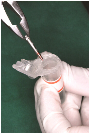
2 × 14 mm stainless steel IZCs (FavAnchorTM SAS, S.H. Pitkar Orthotools Limited, Pune, India) (Figure 1) were placed bilaterally in the IZC area. A total of 36 IZC screws were used in this study. A power analysis was established by G*power, version 3.0.1 (Franz Faul Universitat, Kiel, Germany). With a 95% confidence level and power of test as 80%, where C = 0.5* In[(1 + r)/(1–r)]=0.354, considering the mean difference of 27.9 and standard deviation of 3.01, the calculated sample size came up to 18 per group.
Figure 1. 2 × 14 mm Stainless Steel IZC Bone Screws (FavAnchorTM SAS).
Group I-silver nanoparticle (AgNP) coated (n = 18) and group II-uncoated (n = 18). Pairs of coated and uncoated screws were coded for the right and left sides and random allocation was done to ensure that equal numbers of each type were tested on the right and left sides in a uniform manner.
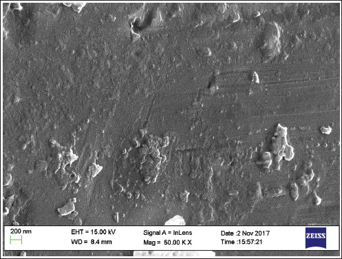
Coating of IZC screws was carried out using magnetron sputtering (Radio Frequency—ANELVA SPF-332H) at CeNSE, IISc-Bengaluru, India. Scanning electron microscope (SEM; JEOL - IT 300, IISc, and Bengaluru, India) with an energy-dispersive spectrometer (EDS) was used to assess the surfaces of the coated IZC screws.

Procedure

The procedure included local anesthesia followed by the placement of the IZC screws, using a Favanchor driver (FavAnchorTM SAS, S.H. Pitkar Orthotools Limited, Pune, India) once the patients were on 0.019″ × 0.025″ SS archwire. IZC screws during initial placement were inserted into the bone perpendicular to the long axis of the adjacent teeth. After penetrating the cortical bone by about 1 mm, the driver was progressively angulated by about 60 to 70 degrees to the maxillary occlusal plane for maximum bone engagement. IZC screws were immediately loaded with a pre-stretched elastomeric chain and reactivated monthly with a distalization force ranging from 8 oz to 14 oz for a period of 6-8 months for correcting class II malocclusion.12 Each screw was routinely evaluated for mobility and ability to maintain continuous anchorage during the treatment period.

Evaluation of Success Rate

Those IZC screws which provided successful anchorage until the end of the IZC screw use were categorized as successful and loose screws that exfoliated or were deemed too loose to provide effective anchorage were categorized under failed screws.

Collection of IZC Screws

All 36 samples of IZC screws were immediately stored after removal at room temperature (15-25°C) and transported in DNAzol transport media for molecular analysis (Figure 2). Followed by terminal-restriction fragment length polymorphism (T-RFLP) analysis in Chromus Biotec Lab, Bengaluru, India. T-RFLP electropherograms were analyzed with Gene Mapper software version 4.1. A Gene mapper was used to determine peak length, height, and area (Figure 3).
Figure 2. Storage and Transport of Retrieved IZC Sample in DNAzol Solution.
Figure 3. Electropherogram Image of Sample No. 7, the X-Axis Marks the Sizes of the Fragments and the Y-Axis Marks the Fluorescence Intensity of Each Fragment.
The numbers of peaks obtained in profiles are an approximate representation of the minimum number of bacteria present in the sample.14 T-RFLP patterns were obtained as table files by sequencing analysis_v 5.4. These sample files contain TRFs (peak values) in base pairs (Figure 4).
Figure 4. Terminal Restriction Fragment Polymorphism (T-RFLP) Data.

Sequencing of the Clones to Identify the Predominant Species

One failed sample each from AgNP coated and the uncoated sample was selected and analyzed in greater detail to identify its nearest species based on 16S rDNA sequence, using the cloning-based identification technique. Five independent clones were sequenced each from AgNP-coated and uncoated samples. The protocol involved PCR amplification, cloning, and sequencing 16S rDNA region from the samples.

Statistical Analysis

Success was modeled as a binomial variable (success-yes/no). Simple descriptive statistics were used to summarize the estimates of success rates across different levels of predictor variables. Each IZC screw was the unit of analysis. Success incidence among groups was analyzed with chi-square analysis. The mean ranks of TRFs among groups were analyzed with Mann-Whitney U test. Data analysis and statistical analysis were performed using SPSS version 20 (IBM Corporation, Chicago, IL, USA) software.

Results

This study was done to compare the success rate of AgNP-coated and uncoated IZC screws.

Nanoparticle Surface Characterization

In this study, 8-10 nm silver nanoparticle coating thickness was obtained by magnetron sputter deposition by optimized data. SEM (Figure 5) and EDS analysis showed that the deposition performed was uniform and successful. EDS spectra plots of the AgNP-coated IZC screws showed the presence of a peak for atomic silver deposited within the surface (Figure 6). Quantification of the spectra indicated that the AgNP-coated IZC screw surface contained 11.89 atomic % of silver (Table 1).
Figure 5. SEM Image of AgNPs Coated IZC Screw.
Figure 6. Energy-Dispersive Spectroscopy (EDS) Image of IZC Screw (A) Uncoated (B) AgNPs Coated.
Table 1. Elemental Composition and Atomic Ordering of AgNP Coated (Table 1A) and Uncoated (Table 1B) Infra-zygomatic Screws as Determined Using Energy-Dispersive Spectroscopy.
AB
ElementWeight%Atomic%ElementWeight%Atomic%
C K11.3741.35O K5.7812.67
Cr K11.089.31Al K41.3053.73
Fe K42.0132.86Cr K10.116.83
Ni K6.184.60Fe K38.2524.04
Ag L29.3611.89Ni K4.562.72
Totals100.00Totals100.00

Success Rate

Of the 18 patients (36 mini-implants) who received IZC screws, the majority were females. Throughout treatment, overall 36.10% of the IZC screws failed. Chi-square analysis inferred that the AgNP-coated IZC screws had a statistically significant higher success rate 77.80% when compared with uncoated IZC screws (50.0%) (P = .023) (Table 2).
Table 2. Association of AgNP Coated and Uncoated Infra-zygomatic Screws with Their Success Rates.
 InferenceTotalP value
FailureSuccess
AgNP CoatedCount41418.023*
% within group22.20%77.80%100.00%
UncoatedCount9918
% within group50.00%50.00%100.00%
TotalCount132336
% within group36.10%63.90%100.00%

Note: *Statistical significance set at .05.

Binary logistic regression analysis shows no statistically significant association between age, sex, and site with success and failure of AgNP-coated and uncoated IZC screws (Tables 3 and 4).
Table 3. Bivariate Analysis in AgNP Coated Infra-zygomatic Screws.
Variables in the Equation
 BS.E.WalddfSig.Exp(B)/ Odd’s Ratio
Step 1aAge.269.340.6251.4291.309
Sex(1).2231.358.0271.8701.250
Site(1)–1.0281.244.6831.409.358
Constant–3.1825.823.2991.585.041

Abbreviations: SEX(1) = Male; SITE(1) = LEFT.

Table 4. Bivariate Analysis in Uncoated Infra-zygomatic Screws.
Variables in the Equation
 BS.E.WalddfSig.Exp(B)/ Odd’s Ratio
Step 1aAGE10.0183237.634.0001.99822430.975
SEX(1)–31.36630729.889.0001.999.000
SITE(1)18.6506475.268.0001.99125787162.775
Constant–180.32858277.410.0001.998.000

Abbreviations: SEX(1) = Male; SITE(1) = LEFT.

Molecular Analysis

The result of T-RFLP profiling is a graph called electropherogram (Figure 3) and the X-axis marks the sizes of the fragments while the Y-axis marks the fluorescence intensity of each fragment. In a T-RFLP profile each peak assumingly corresponds to one genetic variant in the original sample while its height or area corresponds to its relative abundance in the specific community14 (Figure 4).
The mean rank of TRFs (ie species) for the AgNP-coated group was 14.92% and the uncoated group was 22.08%. This difference was statistically significant (P < .05) (Table 5).
Table 5. Comparison of Mean TRF Values Between Coated and Uncoated Infra-zygomatic Screws.
 GroupsNMeanStd. DeviationMean RankZAsymp. Sig. (2-Tailed)
TRF_ValuesCoated Samples1878.6950.2214.92–2.05.04*
Uncoated Samples1890.5741.7022.08
Total3684.6345.89 

Note: *Statistical significance set at .05.

Cloning and Sequencing of the Clones to Identify the Predominant Species

In the present study sequencing of the clones was done to identify the predominant species in the samples. Here we could identify predominant bacteria which were varying between 2 groups of samples. Predominant species in AgNP coated were Prevotella oris strain NCTC13071, Uncultured prevotella sp, and uncultured bacterium, while Capnocytophaga leadbetteri, Capnocytophaga sp. Uncultured bacterium clone, Uncultured bacterium clone 069096_35 were predominant in the uncoated sample (Table 6).
Table 6. Microflora Associated with AgNP Coated and Uncoated Infra-zygomatic Screws.
S. No.Sample: A-7(AgNP Coated)Sample: A-21 (Uncoated)
1Prevotella oris strain NCTC13071Capnocytophaga leadbetteri
2Uncultured Prevotella sp.Capnocytophaga sp.
3Uncultured Prevotella sp. cloneCapnocytophaga sp.
4Uncultured bacteriumUncultured bacterium clone
5Uncultured bacteriumUncultured bacterium clone 069096_353

Discussion

In the present study, silver nanoparticles were deposited onto the surface of 2×14 mm SS IZC screws using Radio Frequency magnetron sputtering. To increase the adhesive strength post-deposition heat treatment was done as AgNP particles were deposited onto a smooth, machined SS surface where adhesion was a big concern. 15 The EDS microanalysis confirmed that a higher atomic % (11.89%) of AgNPs was obtained by magnetron sputter coating.
During the clinical procedure, the IZC screws were initially autoclaved15 and insertion was done under local anesthesia following which immediately loaded with an e-chain. The findings of the present study showed an overall success rate of 63.9% which was slightly lower when compared to previous studies by Chang et al.12 and Uribe et al.16 who had reported 78.2% and 93% success rates, respectively.
The stability of these IZC screws seems to be affected by bone stiffness and bone density.17 Most of the patients in this study were in the growing age group. Chen et al.18 reported that screw stability primarily depends upon the mechanical interlock between the alveolar bone and the screw. Therefore, cortical bone density and thickness are important factors in the success of IZC screws.
The split-mouth design success rate for AgNP coated and uncoated groups were 77.80% and 50%, respectively. A key advantage of this split-mouth design is that the sample size requirement is approximately half that of a parallel-group design when all other parameters are equal. This is because each patient acts as his/her control, so much of the inter subject variability is removed, resulting in increased study power or a decrease in the number of participants required compared with a study in which patients receive only one intervention.19 The higher success rate for the AgNP-coated group when compared to the uncoated group may be mainly due to the antibacterial effect of AgNPs. This nanometal attaches to the surface of the bacterial cell membrane and drastically disturbs its permeability and respiration and thus has high antibacterial actions.20 Binary logistic regression analysis shows no statistically significant association between age, sex, and site with success and failure of AgNP-coated and uncoated IZC screws. However, the present study sample size is small and statistically insignificant to evaluate and confirm the same.
This is probably the first in-vivo study that compared the efficacy of AgNPs coated and uncoated SS IZC screws, whereas the previous study was in-vitro and has shown similar findings.
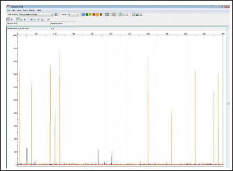
6
T-RFLP analysis, a DNA-based nonculture-dependent approach was used in this study for molecular analysis. This approach helped in the rapid assessment of the bacterial colonization around the AgNP-coated and uncoated IZC screw surfaces.14 In the study TRFs detected per sample ranged from 2 to 12 in group I and 4 to 15 in group II. Among these, the TRF with a length of 107 base pair (briefly TRF107) was the most frequently encountered (88.9%) across samples followed by TRF103(86%), TRF109(80.5%), TRF80(66.7%), TRF365(63.9%), TRF55(55.5%) and TRF454(41.7%). These values indicate that TRFs were statistically significantly distributed between the 2 groups.
TRF107 and 103 were the most frequent species in the uncoated group while in the coated group TRF107 and 103 were counted 15 times (out of 18). This indicates TRF107 and TRF103 were reproducibly present across 2 groups signifying the reproducibility and sensitivity of the T-RFLP protocol followed in the present study. T-RFLP data analysis results also showed that few peaks were present only in uncoated samples. These peaks included TRF122, TRF124, TRF125, TRF157, TRF144, TRF229,TRF237, TRF256,TRF254, TRF457,TRF278,TRF399.TRF433,TRF445,TRF255,TRF261,TRF239,TRF432,TRF453. Also, a few other peaks were found only in AgNP coated group which included TRF461, TRF460, TRF457, TRF478, TRF246, TRF271, and TRF272. The result suggests that the protocol was sensitive enough to identify the peaks.
The T-RFLP analysis in the present study revealed a statistically significant difference in the TRF value between AgNP-coated and uncoated IZC screws. The mean rank of TR-Fs was lower in AgNP coated group (14.92%) compared to the uncoated group (22.08%). The reason may be with higher atomic % of AgNP deposition on the surface of IZC screws. This finding was similar to an in-vitro study by Venugopal et al.6 who concluded that the biopolymer modified AgNP coated titanium screws with higher atomic% exhibited greater antibacterial effects with clear zones of inhibition.
PCR-cloning sequencing protocol was used in the present study to identify the predominant species within the microbial population present between a coated and uncoated sample. Prevotella oris strain NCTC13071, Uncultured prevotella sp, and uncultured bacterium were predominant species in failed AgNP coated sample, while Capnocytophaga leadbetteri, Capnocytophaga sp. Uncultured bacterium clone, Uncultured bacterium clone 069096_35 was the predominant species in the failed uncoated sample.
Here the term “unculturable bacteria” is used to denote uncharacterized organisms that cause oral infections.21 The predominant species detected in the present study might have been overlooked in an in-vitro culture as 50% of the oral flora is unculturable. The nonculturability in vitro may be due to the disruption of bacterial cytokine networks. Bacterial cytokines are thought to be mediators of bacterial signaling and may be particularly important in coordinating the growth of component organisms in bacterial biofilms such as dental plaque. 22
This underlines the usefulness of nonculture-dependent molecular techniques (16S rDNA PCR and sequencing) used in the present research for studying samples. Apart from that, the predominant species identified in failed samples, both AgNP coated and uncoated, were found to be gram-negative. This result was found to be similar to that described in the earlier report by Kumar et al.23 wherein using pyrosequencing, the investigators had confirmed that the failed IZC screws were dominated by gram-negative bacteria. AgNPs were found to have a stronger antibacterial effect on gram-negative bacteria compared to gram-positive bacteria.8 Thus AgNPs incorporation around IZC screws would impart stronger antibacterial efficacy ensuring long-term stability and success.
In future studies more elaborate protocols of the Next Generation Sequencing based methodology may be employed. Through that we may effectively sequence and identify the complete metagenome of all organisms present. This would effectively find all minor species present or absent within samples. Also, the advantages of biogenic metallic nanoparticles over physio-chemically obtained nanoparticles may be considered for future research.
We had considered cases with congenitally missing third molars or samples where third molars were extracted before the orthodontic treatment in our study but standardization of the cases would be necessary to avoid selection bias in studying the antibacterial efficacy of AgNPs coated and uncoated IZC screws. This would be considered a limitation of our study.

Conclusion

In this study, 8 to 10 nm silver nanoparticle deposition was obtained by magnetron sputter coating. The EDS microanalysis confirmed that 11.89 atomic % of AgNPs was deposited over the IZC screw surface.
The study implemented a split-mouth experimental design to compare the in-vivo efficacy of AgNPs coated and uncoated IZC screws. The success rate with AgNP-coated IZC screws was significantly higher than the uncoated screws.
Predominant bacteria were found to be gram-negative, which were varying between failed AgNP coated and uncoated samples and which need to be explored subsequently.

Ethical Approval

The study was approved by the Institutional Ethical Committee (No:110/2017-18).

Informed Consent

The participant has consented to the submission of the article to the journal.

Declaration of Conflicting Interests

The authors declared no potential conflicts of interest with respect to the research, authorship, and/or publication of this article.

Funding

The authors received no financial support for the research, authorship, and/or publication of this article.

ORCID iD

References

1. Ghosh A. Infra-zygomatic crest and buccal shelf-orthodontic bone screws: a leap ahead of micro-implants—Clinical perspectives. J Indian Orthod Soc. 2018s;52:215–223.
2. Zitzmann NU, Berglundh T. Definition and prevalence of peri-implant diseases. J Clin Periodontol. 2008;35(8 Suppl):286–291.
3. Sato R, Sato T, Takahashi I, Sugawara J, Takahashi N. Profiling of bacterial flora in crevices around titanium orthodontic anchor plates. Clin Oral Implants Res. 2007;18(1):21–26.
4. De Freitas AO, Alviano CS, Alviano DS, Siqueira JF Jr, Nojima LI, Nojima Mda C. Microbial colonization in orthodontic mini-implants. Braz Dent J. 2012; 23(4): 422–427.
5. Mombelli A, van Oosten MA, Schurch E Jr, Land NP. The microbiota associated with successful or failing osseointegrated titanium implants. Oral Microbiol Immunol. 1987;2(4):145–151.
6. Venugopal A, Muthuchamy N, Tejani H, Gopalan AI, Lee KP, Lee HJ, Kyung HM. Incorporation of silver nanoparticles on the surface of orthodontic microimplants to achieve antimicrobial properties. Korean J Orthod. 2017;47(1):3–10.
7. Roy A, Bulut O, Some S, Mandal AK, Yilmaz MD. Green synthesis of silver nanoparticles: biomolecule-nanoparticle organizations targeting antimicrobial activity. RSC Adv. 2019;9:2673–702.
8. Qing Y, Cheng L, Li R, Liu G, Zhang Y, Tang X, Wang J, Liu H, Qin Y. Potential antibacterial mechanism of silver nanoparticles and the optimization of orthopedic implants by advanced modification technologies. Int J Nanomedicine. 2018;13:3311–27.
9. Gilani RA, Laxmikanth SM, Ramachandra CS, Prasad SL, Shetty S, Vasudevan SD. Antibacterial and antiadherent properties of silver dioxide-coated brackets. J Indian Orthod Soc. 2017;51:9–14.
10. Ghasemi T, Arash V, Rabiee SM, Rajabnia R, Pourzare A, Rakhshan V. Antimicrobial effect, frictional resistance, and surface roughness of stainless steel orthodontic brackets coated with nanofilms of silver and titanium oxide: a preliminary study. Microsc Res Tech. 2017;80(6):599–607.
11. Moreira DM, Oei J, Rawls HR, Wagner J, Chu L, Li Y, Zhang W, Whang K. A novel antimicrobial orthodontic band cement with in situ-generated silver nanoparticles. Angle Orthod. 2015;85(2):175–183.
12. Chang CH, Lin JS, Roberts WE. Failure rates for stainless steel versus titanium alloy infrazygomatic crest bone screws: a single-center, randomized double-blind clinical trial. Angle Orthod. 2019;89(1):40–46.
13. Marchi A, Camporesi M, Festa M, Salvatierra L, Izadi S, Farronato G. Drilling Capability of Orthodontic Miniscrews: In Vitro Study. Dent J. 2020;8:138
14. Hommez GM, Verhelst R, Vaneechoutte M, Claeys G, De Moor RJ. Terminal restriction fragment length polymorphism analysis of the microflora in necrotic teeth of patients irradiated in the head and neck region. J Endod. 2008;34:1048–1051.
15. Juhasz JA, Best SM. Surface modification of biomaterials by calcium phosphate deposition. In: Williams R, ed. Surface Modification of Biomaterials: Methods Analysis and Applications. 1st ed. Woodhead Publishing Series in Biomaterials; 2011:143–169.
16. Uribe F, Mehr R, Mathur A, Janakiraman N, Allareddy V. Failure rates of mini-implants placed in the infrazygomatic region. Prog Orthod. 2015;16:31.
17. Swami V, Vijayaraghavan V, Swami V. Current trends to measure implant stability. J Indian Prosthodont Soc. 2016;16(2):124–130.
18. Chen YJ, Chang HH, Huang CY, Hung HC, Lai EH, Yao CC. A retrospective analysis of the failure rate of three different orthodontic skeletal anchorage systems. Clin Oral Implants Res. 2007;18(6):768–775.
19. Hujoel PP, DeRouen TA. Validity issues in split-mouth trials. J Clin Periodontol. 1992;19(9):625–627.
20. Ramasamy M, Lee J. Recent nanotechnology approaches for prevention and treatment of biofilm-associated infections on medical devices. Biomed Res Int. 2016; 2016:1851242.
21. Stewart EJ. Growing unculturable bacteria. J Bacteriol. 2012;194(16):4151–4160.
22. Wade NW. Unculturable bacteria–the uncharacterized organisms that cause oral infections. J R Soc Med. 2002;95(2):81–83.
23. Kumar PS, Mason MR, Brooker MR, O’Brien K. Pyrosequencing reveals unique microbial signatures associated with healthy and failing dental implants. J Clin Periodontol. 2012;39(5):425–433.