

Spider silk enhanced tissue engineering of cartilage tissue: Approach of a novel bioreactor model using adipose derived stromal cells
Abstract
Introduction
Methods
Cell culture
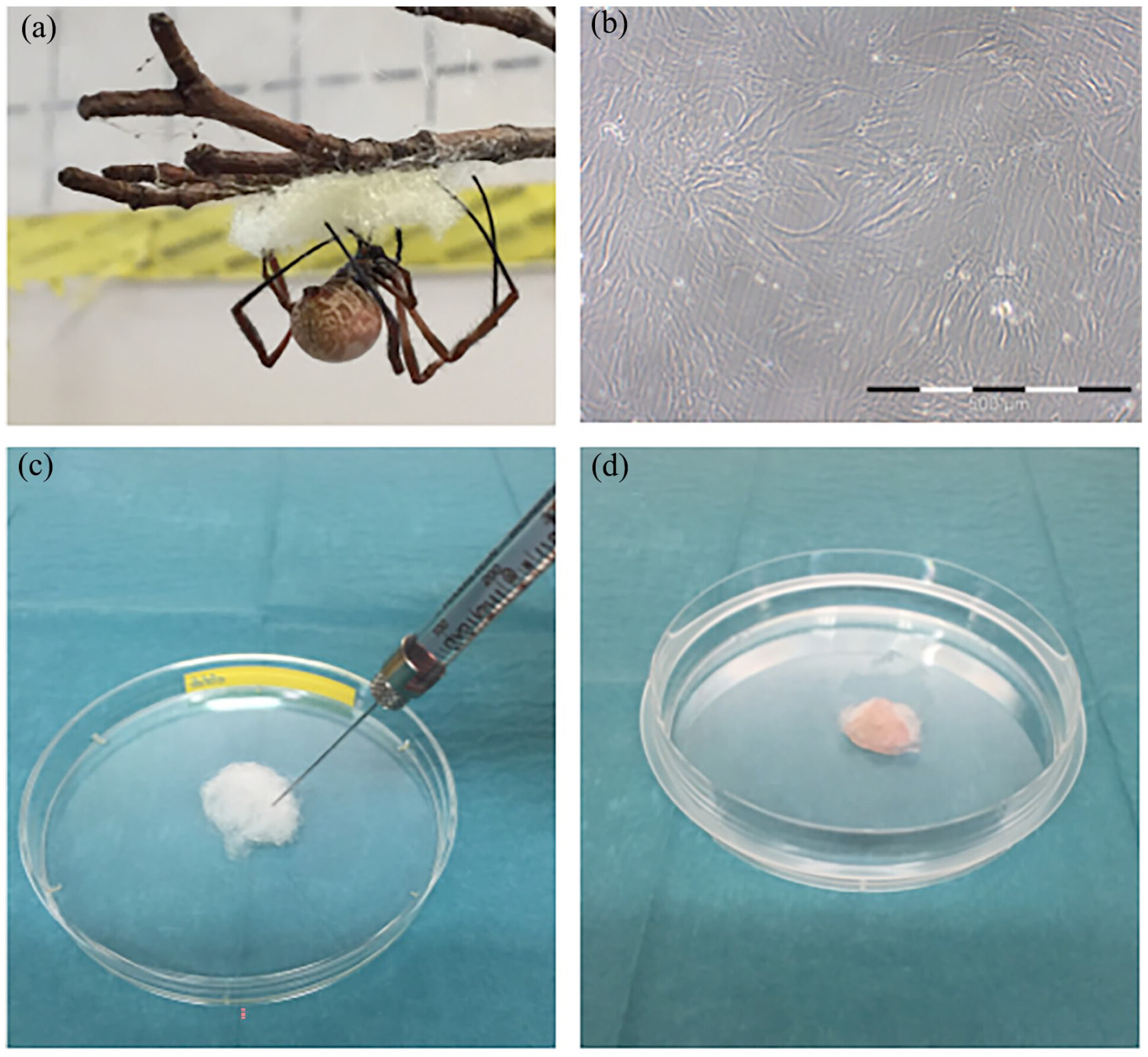
Animal handling and harvesting procedure of spider silk
Construct preparation
| Experimental group number | Type of differentiation | Number of samples |
|---|---|---|
| 1 | Chemical (TGF-β2 and BMP-7) | (n = 8) |
| 2 | Mechanical (bioreactor) | (n = 8) |
| 3 | Mechanical-chemical (bioreactor + TGF-β2 and BMP-7) | (n = 8) |
| 4 | None (controls) | (n = 8) |
Chemical induction of differentiation
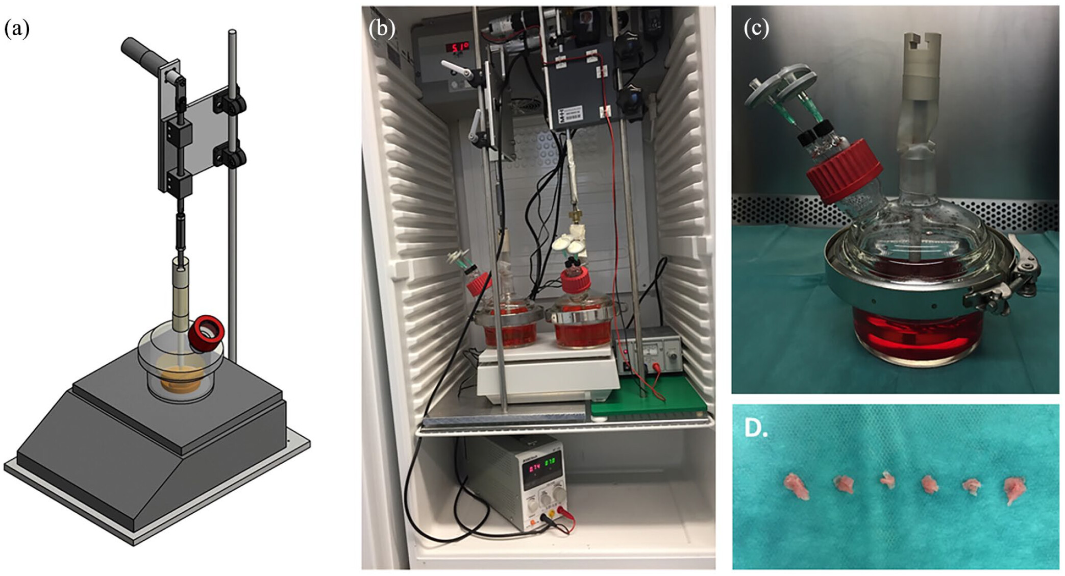
Concept of bioreactor cultivation and mechanical induction of differentiation
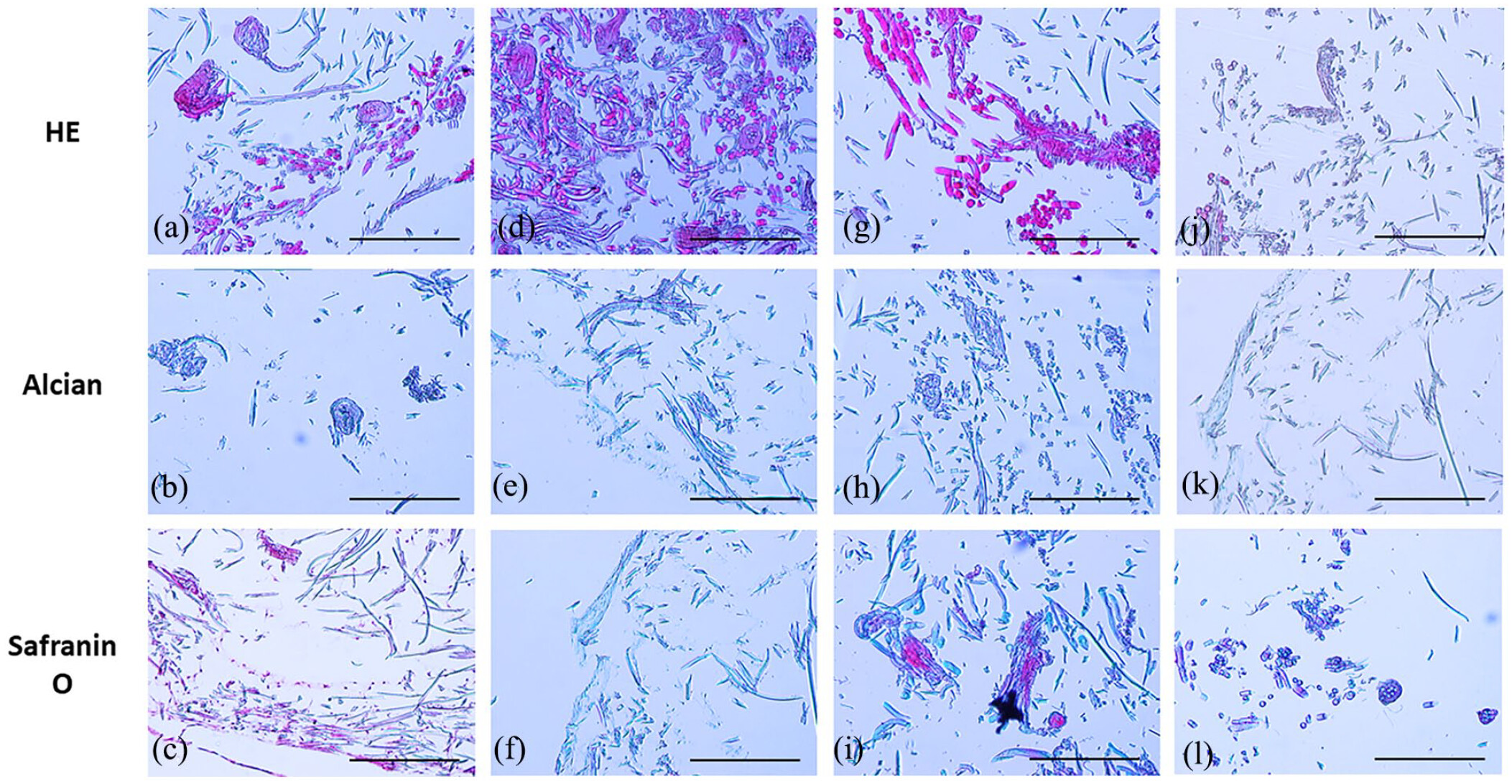
Histology
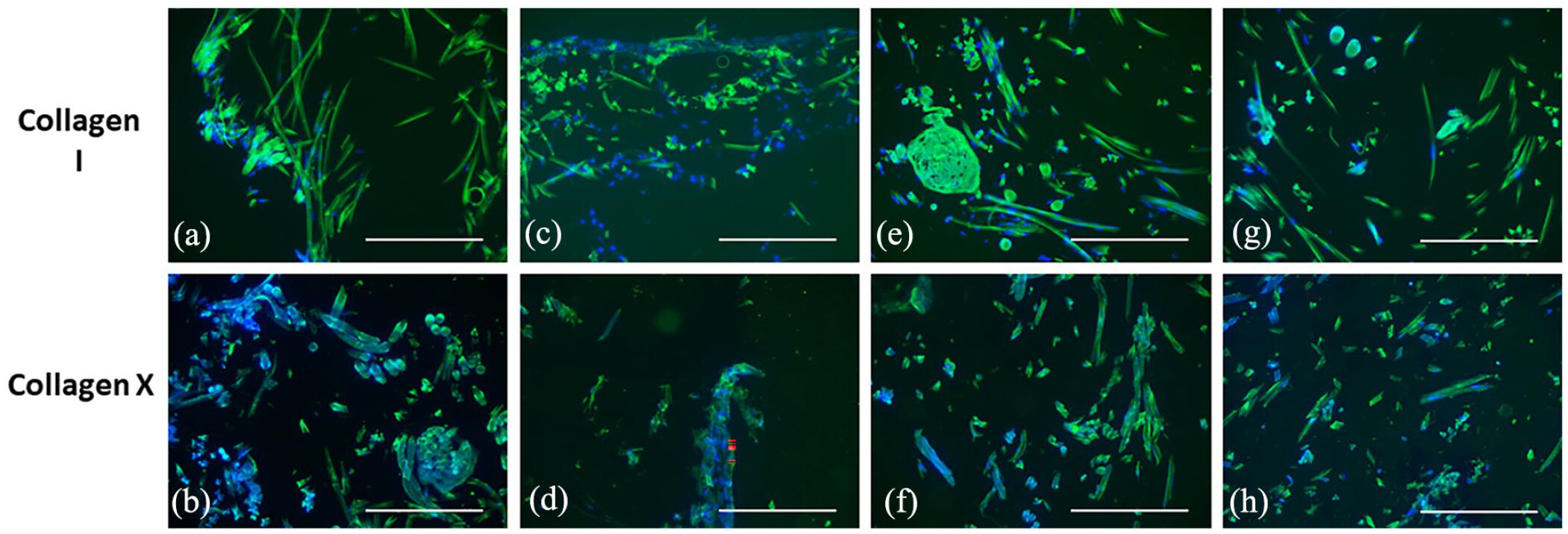
Immunohistochemical staining
Results
Successful preparation of densely seeded silk constructs
Chemical induction of chondrocyte differentiation

Design and construction of the bioreactor for mechanical induction of differentiation
Histological analysis showed a change in cell morphology and first evidence for ECM production
Immunohistochemical stainings indicated chondrogenic differentiation of rASC

Discussion
Conclusions
Acknowledgments
Declaration of conflicting interests
Funding
ORCID iD
Footnote
References
Cite
Cite
Cite
Download to reference manager
If you have citation software installed, you can download citation data to the citation manager of your choice
Information, rights and permissions
Information
Published In
Keywords
Rights and permissions
Authors
Contributorship
Metrics and citations
Metrics
Journals metrics
This article was published in Journal of Applied Biomaterials & Functional Materials.
View All Journal MetricsPublication usage*
Total views and downloads: 1560
*Publication usage tracking started in December 2016
Publications citing this one
Receive email alerts when this publication is cited
Web of Science: 7 view articles Opens in new tab
Crossref: 6
- Application of Spider Silk-Based Materials in Regenerative Medicine: From In Vivo Studies to Clinical Use
- Advancing Cartilage Tissue Engineering: A Review of 3D Bioprinting Approaches and Bioink Properties
- Delivery Strategies of Growth Factors in Cartilage Tissue Engineering
- Exploring the Unique Properties and Superior Schwann Cell Guiding Abilities of Spider Egg Sac Silk
- Mechanical and Biological Characterization of Ionic and Photo-Crosslinking Effects on Gelatin-Based Hydrogel for Cartilage Tissue Engineering Applications
- “Unravelling the mysteries of spider silk: A comprehensive review of its properties and applications”
Figures and tables
Figures & Media
Tables
View Options
View options
PDF/EPUB
View PDF/EPUBAccess options
If you have access to journal content via a personal subscription, university, library, employer or society, select from the options below:
I am signed in as:
View my profileSign out

I can access personal subscriptions, purchases, paired institutional access and free tools such as favourite journals, email alerts and saved searches.
loading institutional access options
Alternatively, view purchase options below:
Purchase 24 hour online access to view and download content.
Access journal content via a DeepDyve subscription or find out more about this option.
