Intended for healthcare professionals
Free access
Research article
First published online November 22, 2023

Mineralized Collagen Fiber-based Dental Implant: Novel Perspectives

Abstract

Aim:

One of the problems that dental bone defects commonly face is less biocompatibility. Therefore, it is important to find effective natural dental materials to increase the rate of cell viability. In the present work, a blend of mineralized collagen fiber (MCF)/silica nanoparticles (Si-NPs) is used to develop a dental implant (DI), for their possible application in dental bone repair.

Materials and Methods:

This research study was to develop a technique for the fabrication of DI using natural materials. Accordingly, the present work provides DI, essentially by PVA (6g): MCF (1.5g): Si-NPs (0.8g): CaCO3 (1.0g) slurry into DI with other conventional implants. The DI was characterized by its mechanical, physicochemical, and biocompatibility study. The mechanical analysis was statistically different in all three time periods (p < .05). Surface characterization of DI was carried out before and after immersion in the SBF.

Results:

The DI was excellent mechanical properties like compressive strength (24.22 ± 0.32 MPa) and elongation at break (16.51% ± 0.71%). The morphology of the DI showed a good pore size observed. Bioactivity test was observed on the Calcium/Phosphate of the DI. The biocompatibility of the study MTT (3-(4,5-dimethyl) thiazol-2-yl-2,5-dimethyl tetrazolium bromide) assay using the MG63 (human osteoblast cell line) has proven to more viable cell on the DI.

Conclusion:

The study has devised a process for using fish waste in the preparation of DI. The DI with the required strength, biocompatibility, and bone mineralization properties may be tried as a DI in large animals after obtaining the necessary approval.

Introduction

The most prevalent protein with fibrous structure in all organisms, collagen (C), serves as the primary functional component of extracellular structures in tendons and ligaments. Glycine is found in every third location of the proteins considerably formed themselves triple-helix construction, which is arranged helicoidally. The substances from which collagen molecules are extracted and the post-processing conditions can change the crystalline structure and molecular features, particularly their ability to interact with the derived components. Recently, collagen fibers are being widely explored due to the growing interest in novel biomaterials enhanced with molecules of collagen for a variety of biological purposes.1 Among the many biological sources of collagen used for health advantages, bovine skin, tendons, and intestines are potential candidates for widespread use as biomedicine. Collagen-based polymers have been employed in both pharmaceuticals and healthcare. Nearly every physical form, including liquids, gels, powders, fibers, membranes, sponges, tubes, etc., contains collagen.
Fracture healing is a complex biological phenomenon that involves the restoration of structural integrity through bone regrowth. Losing a tooth can be distressing on the mind. Replacement attempts for missing teeth were performed even in primitive times. Implant dentistry unique is in its ability to achieve this goal despite stomatognathic system atrophy, disease, or damage. To achieve the optimum objectives of implant dentistry, the hard and soft tissues must have appropriate volumes and attributes. If there is not enough bone, a variety of surgical methods can be used to reconstruct the weak ridge for implant implantation.2 Bone tissue engineering technologies have been explored for a variety of dental applications such as salivary gland regeneration, tooth development, and pulp dentin complex regeneration, with promising results.3 Javed et al.4 report the bone growth factors have a significant impact on protein synthesis, chemotaxis, cellular proliferation, and differentiation. Non-collagenous proteins found in demineralized dentin and bone matrices include dentin matrix protein-1, dentin sialophosphoprotein (DSPP), osteonectin, osteopontin, and bone sialoprotein. Bone morphogenetic proteins (BMPs) and fibroblast growth factors are also found in demineralized matrices.5
Bone manipulation methods can affect the density of a person’s bones to improve their tensile strength and durability. These methods mobilize necessary bone through the plastic bending, shaping, or condensing of tissue into a bone flap or bone-periosteal flap. These change the size or shape of the bone while keeping it healthy and whole. The goal is to modify the residual bone to create an intra-body chamber that heals similarly to an extraction site and allows mesenchyme stem cell accessibility in addition to normal wound healing mechanisms. Choosing methods for ridge manipulation and the form of the bone defect could be taken into account. The main drawback of synthetic implants is their substantial modulus of elasticity, which results in a stress-shielding impact that increases bone resorption since cortical bone’s elastic modulus is incongruent with titanium’s elastic modulus.6 Fractures and faults may already exist, or they may develop as a result of the implant. Blood transfusions are performed in the human body more frequently than bone transplants. Therefore, it is essential to carefully consider orthopedic device design to protect patients and efficiently treat bones and joint disorders.7
The parameters of design and choice of material for bone implants are determined by functional and biomechanical requirements. To achieve biocompatibility, a variety of biomaterials are used, including metals, ceramics, polymers, and composites. Biomedical implants can take the role of worn-out organs, joints, and tissues. For them to achieve integrity, according to International Standards Organization (ISO) 20160, a homogeneous and stable microstructure is required. An ideal implant should be able to work properly without requiring any more revision surgery. The majority of orthopedic biomedical implants are used for hip, knee, spinal, and maxillofacial replacements.8
Dental implant (DI) technology has progressed in the past few years, providing patients with unparalleled levels of affordability, convenience, and effectiveness. This is one of the main reasons why so many dentists believe that DIs are the best approach to replace missing teeth. Natural-based DIs can provide numerous advantages.9 Previously, the researchers considered a natural-based implant in tissue engineering and dental surgery while focusing on cost-effective techniques. The main aim of this research work focused on the preparation of natural material-based DIs and evaluated using the mechanical, physicochemical, and biological properties.

Materials and Method

Preparation of Mineralized Collagen Fibers (MCF)

The taxonomic status of the Indian fish (Caranx melampygus) was determined and validated using 500 grams of raw fish bone. The MCF, which served as the starting material, was successfully isolated. The fishbone was treated with sodium salt (1 wt%) and carefully cleaned before being stored at 4°C until future usage. The sodium salt-treated bone (size 2 cm × 2 cm) and cleaned debris with water and dried at 27°C for 12 h. The dried samples were then defatted using chloroform: acetone (ratio 3:2) and then mineralized using 1 N HCL for 7 days at 6°C. The mineralized materials were dried using an oven for 3 h at 35°C to get the fibrous samples converted into MCF using a retch grinder machine.

Extraction of Rhus Coriaria L. Extract

Rhus coriaria L. seeds were gathered and dried at 25°C in the shade, followed by being ground in a grinder. 40 g of crushed Rhus coriaria L. was heated with 80 mL of deionized water using a magnetic stirrer. After filtering the final combination through the filter paper, the pure extract was kept at 8°C for later use.

Preparation of Silica Nanoparticles (Si-NPs)

20 mL of Na2SiO3 was added to 100 mL of Rhus coriaria L. extract, which was then stirred at 60°C. This process was carried out under the influence of reflux for 12 h at pH 9 and before the development of the SiO2. The solution pH was changed using NaOH, which was thoroughly dissolved in 50 mL of deionized water. As a result, the mixture was filtered, and impurities were completely washed from the precipitate with methanol and water. Centrifugation at 7000 rpm for 25 min separated the mixture from the leftovers; the contaminants and organic elements surrounding the Si-NPs were then eliminated by heating the mixture to 550°C.

Preparation of Dental Implant

6 g of PVA was added to 100 mL of deionized water to generate 6% (w/w) PVA glue. The mixture was then heated in a water bath at 6°C for 4 h until the PVA entirely dissolved. The 100 mL of PVA glue was simultaneously mixed with 1.5 g of mineralized collagen and 0.8 g of Si-NPs. The bioceramic implant was then prepared by adding 1 g of calcium carbonate powder and solid mixing it for 10 mins. Bioceramic was prepared and used to design the implant dimensions according to the method used in our previous work.10 In brief, DI were prepared by blending PVA glue, mineralized collagen, and Si-NPs with the help of using rotary mixer (MIXYVAC S, Italy), and the resultant mixture was loaded into the injection molding system (AV-YD Denture Injection system, Aixin Medical Equipment Co., Ltd. China) and pulled out with an appropriate glass rod. The produced DI was dried at 55°C at 70°C for 12 h. DI had a length of 2 to 4 cm, with an average diameter of 2–10 mm.

Characterization

The surface morphology and microstructure of DIs were observed by a high-resolution scanning electron microscope (HR-SEM) ((LEICA Stereoscan 440)). The chemical composition of the bioceramic was determined by Fourier transform infrared spectroscopy (FTIR) (Nicolet 360). The mechanical features were measured using INSTRON (1405) equipment. The level of biocompatibility was evaluated using the MTT method. The bioactivity of the implants was assessed by immersing them in SBF, which has an ionic structure similar to that of human blood plasma. The simulated bodily fluid (SBF) solution was prepared according to the literature.11 The DI was immersed in the SBF solution for 7 days. After the seventh day, the implant was removed and cleaned with distilled water and HRSEM. The occurrence of phosphate and calcium on the outer layer of DI was confirmed using energy-dispersive X-ray analysis (EDX).

Results

Characterization of MCF and Si-NPs

FTIR spectroscopy was determining the structural configuration of a chemical molecule through the absorption peak of the distinctive groups. In FTIR spectroscopy, amide I (1634 cm–1), amide II (1552 cm–1), and amide III (1244 cm–1) are the absorption peaks of the traditional collagen structure (Figure 1a). In Figure 1b, the HRSEM was displayed. The micro-spatial network structure of the mineralized collagen was observed using HRSEM with various microstructures. This structure has continuous and discontinuous portions that were arranged in a particle-like manner. The EDX spectrums of demineralized collagen are presented in Figure 1c. The result showed the samples were observed Calcium and phosphate content.
Figure 1. (a) FTIR Spectra of Si-NPs, (b) HRSEM Image of Si-NPs, and (c) EDX Spectra of Si-NPs.
FTIR spectrum of Si-NPs is shown in Figure 2a. Two prominent peaks were observed in the spectra at around 1053 cm–1 and 786 cm–1. These peaks correspond to the asymmetric vibration of stretching of the siloxane link (Si-O-Si) and the symmetric vibration of the Si–O bond, respectively. HR-SEM was analyzed the Si-NPs surface morphology are displayed in Figure 2b. It is evident that most of the Si-NPs are spherical and nanoscale in size. The Si-NPs have a certain degree of agglomeration, which is typical of the green synthesis process. The elemental identification of the Si-NPs by EDX spectrum is shown in Figure 2c. These findings demonstrate the capability of the Si-NPs with sizes below 20 nm.
Figure 2. (a) FTIR Spectra of MCF, (b) HRSEM Image of MCF, and (c) EDX Spectra of MCF.

Characterization of DIs

The DI functional structures are shown by FTIR (Figure 3a). The value observed at 3280 cm–1 is for the asymmetric bending vibration of CH2, the peak at 1630 cm–1 is for the vibration of stretching of C=O, the peak at 1545 cm–1 is for the vibration of bending peak of N–H and C–N, and the peak at 1235 cm–1 is for the CH2 movement of the proline and glycine sections from the chains of peptides. The DI SEM pictures are depicted in Figure 3b. We identified that the DI had an even surface with several equally sized pores that were around the same size. It was discovered that the DI’s pores had an average diameter of 355 µm. The Si-NPs are equally dispersed throughout the DI matrix and are roughly of the same shape. The non-agglomerated regions’ average Si-NPs particle size ranges from 20 to 40 nm. DI has a porous, mineralized, fibrous mesh-like appearance in SEM images, which suggests that the material can facilitate wound exudate absorption, proliferation, cell attachment, and migration for tissue regeneration, as well as the exchange of air and oxygen diffusion at the wound site. These characteristics are crucial for bone healing dressing materials. The DI possessed the compressive strength of about 24.22 ± 0.32 MPa and elongation at break value of 16.51 ± 0.71.

Bioactivity Test of DI

Figure 3c and 3d shows the HRSEM and EDX for DI following 7 days of SBF immersion. It was discovered that the Ca/P ratio is the same as the hydroxyapatite ratio. Results with DI immersed in SBF solution have demonstrated that an unintentional calcium phosphate layer forms on the implant surface.
Figure 3. (a) FTIR Spectra of DI, (b) HRSEM Image of DI, (c) HRSEM Image of DI After SBF Treatment, and (d) EDX Analysis of DI After SBF Treatment.

Biocompatibility Study

The biocompatibility of the developed DI was assessed based on the rate of cell growth using MG63 cells and the MTT assay. The MG63 cells could be shown to keep growing on the implant’s surface, proving that fibroblast cells weren’t harmed by the implant. The results showed that DIs are significantly greater rate of cell proliferation compared to a control group. The level of cell growth was reported to be practically identical in DI and the control group on 6 h and day 1 of post-seeding; however, DI demonstrated a dependent on time improvement in cell growth at days 1, 2, and 3 of post-seeding (Figure 4a). According to fluorescence microscopic images of Figure 4b, the quantity of growth cells was greatly higher in the DI displayed group than in the controls.
Figure 4. (a) MTT Assay Demonstration of Control Sample and DI on MG-63 Cells, (b) Fluorescence Micrographs (20X) of MG-63 Cells Cultured on Days 1, 2, and 3.
Note: The asterisks (*) indicate statistically significant differences compared to the control p < .05.

Discussion

Patients who have DIs could be able to talk and eat more effortlessly because their dentures are not at risk of dislodging. Dentures must be replaced when the gum tissue shrinks and the fit changes; implants are not affected by this issue. DIs require less upkeep and hygiene than dentures. Another factor driving the continuous expansion of the global market for DIs is the fact that DIs are an effective treatment for edentulism, as well as the increased desire for cosmetic dentistry across all age groups worldwide.12
Choosing the best bone implantation material from the many choices available is difficult. Although the level of failures and surgical factors must be addressed when selecting a graft material, the collagen-based DI may be offered an alternative when tooth extraction is essential due to its autogenous origin and favorable biocompatibility outcomes. In this study, DI was prepared using natural resources and it was evaluated using bone tissue engineering. The observed quality of DI prepared from MCF and Si-NPs were higher mechanical properties when compared to quality from other implants like Osuchukwu et al.
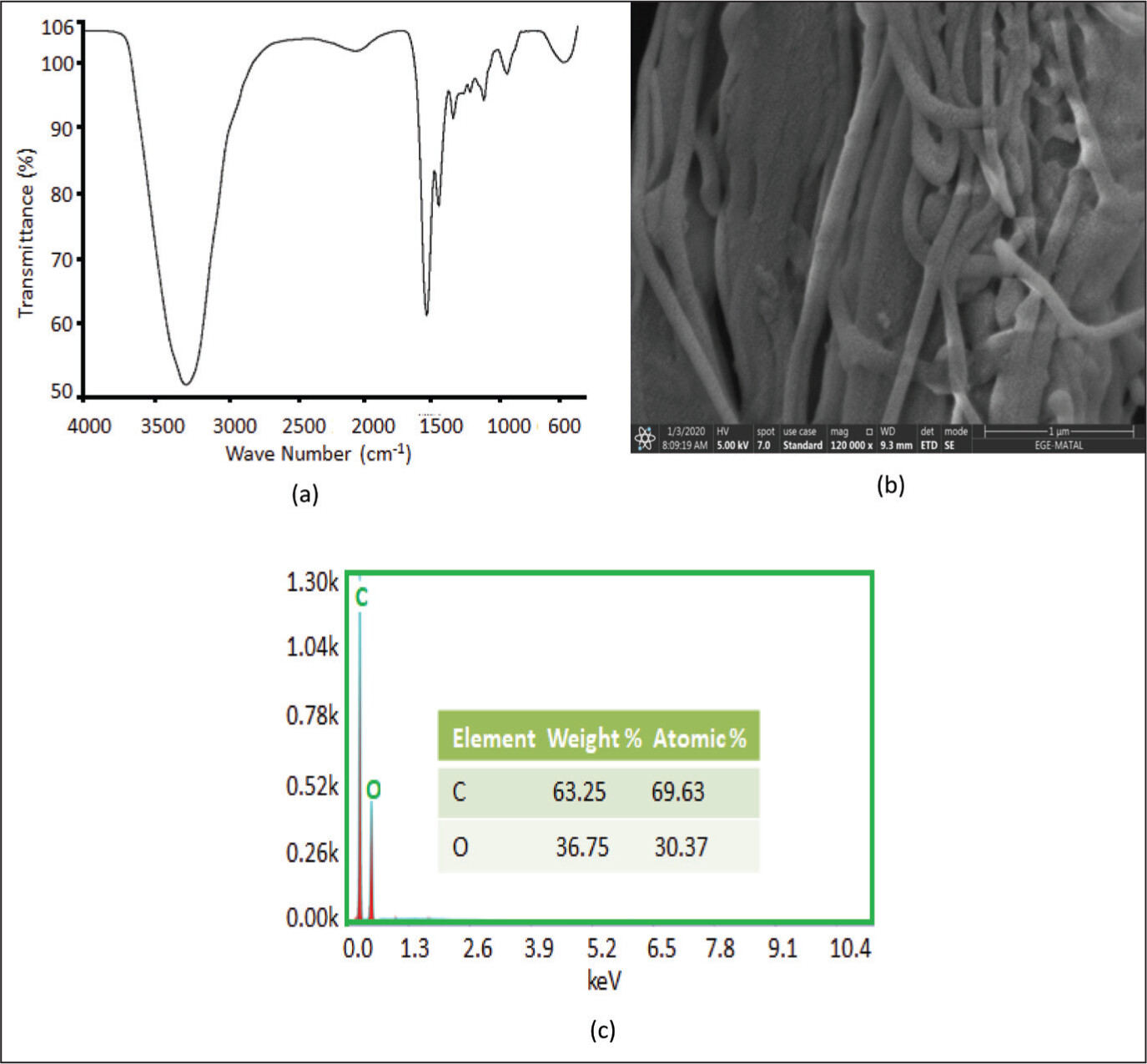
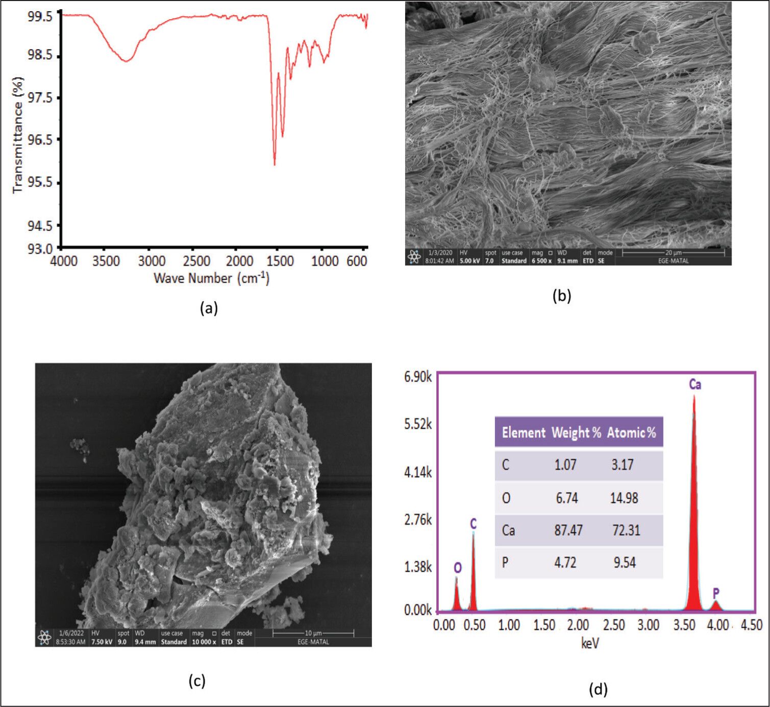
13
Collagen has been found in living organisms as a part of bone, cartilage, and fibrous tissue. More than 90% of the protein in the bone matrix is collagen. The majority of the collagen in bone is Type I collagen, which is produced by osteoblasts. Cross-linking connections are generated between the molecules of collagen in collagen fibrils, which produce interconnected collagen filaments, which develop collagen molecules self-associate into aggregated to form collagen fibrils.14 During the growth, remodeling, and regeneration of bone, osteoblasts produce collagen fibers that are then coated with calcium phosphate, completing the process of building a robust bone matrix.
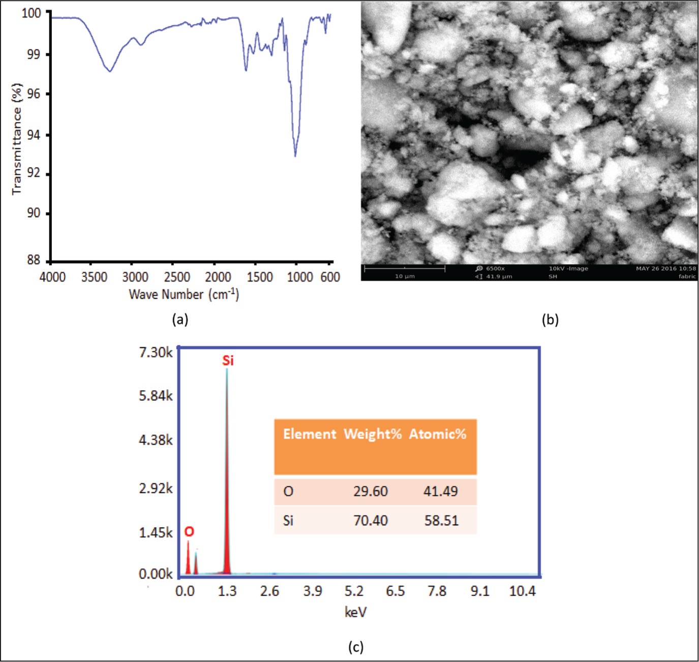
15 Silica nanoparticles (Si-NPs) are the ideal nanomaterials for bone tissue engineering because of their distinctive qualities, including high specific surface area, pore size, multi-functional ability, variable shape, and good biocompatibility.16 Bioactive bone fillers composed of collagen and calcium phosphate have been used in bone repair treatment for many years. They are renowned for their excellent biocompatibility, osteoconductivity, and surgical handling efficiency. However, because of their poor mechanical qualities, such as their extreme brittleness, these bone replacements are frequently not suitable for weight-bearing purposes.17
The presence of these collagen triple helix absorption peaks suggests that the triple helix of structural collagen was mostly retained after mineralization. The P and Ca elements throughout the mineralization process changed the calcium-deficient HA and apatite crystals into amorphous calcium phosphate (ACPs).18 The scissor-like bend vibration of water molecule H2O causes the band at 1637 cm–1. Similar to this, the Si–O and Si–O–Si linkage vibrations’ asymmetric stretching can be seen in the band at 1097 and 802 cm–1, respectively. Additionally, the peak at 463 cm–1 was related to the O–Si–O modes’ bending vibration.19 The Si-NPs result is explained by the greater surface area and persistent affinity of nanoparticles throughout the dehydration process, which results in the agglomeration of the nanoparticles.20 The phytochemicals present in plants and the extraction technique has a major impact on the longevity of NPs and their aggregation into aggregates. The Si-NPs cling together as a result, and they independently produce asymmetrical clusters.21 The measurement efficiency depends on Si-NPs compounds with a low standard number of atoms, like polymers and oxides, making it difficult to identify Si-NPs smaller than 20 nm with modern instruments.22
Hydrogen bonding between collagen, calcium carbonate, and Si-NPs causes the implant to widen and OH vibration of stretching to a lower wave number. It is stated that electrostatic and hydrogen bonding interactions between collagen and the Si-NPs are expected non-covalently.23 The results of the research support the earlier publication about collagen-based bone implants. The collagen-based implant materials need to have enough mechanical strength to handle them and be used in the intended medical applications.24 The expected characteristics from the implants are near to the features of the desired bone in terms of sufficient mechanical strength, modulus of elasticity, corrosion behavior, as well as biocompatibility to prevent adverse effects on humans.25 Design parameters and material selection for bone implants are determined by functional and biomechanical requirements. To achieve biocompatibility, a variety of biomaterials are used, including composites, polymers, ceramics, and metals.26 According to Kokubo and Takadama,27 the development of a phosphate-containing calcium layer on an artificial substance is a crucial step for in vivo bone growth, commonly referred to as bone-like apatite, on the surface of the material. The bone-like apatite seems to trigger signaling proteins and tissues to start the chain of reactions that results in bone formation. DIs’ porous design not only aided in cell migration but also allowed for the transfer of oxygen and nutrients with cells, which is crucial for the formation of the Matrix and vascularization during bone tissue regeneration.28

Conclusion

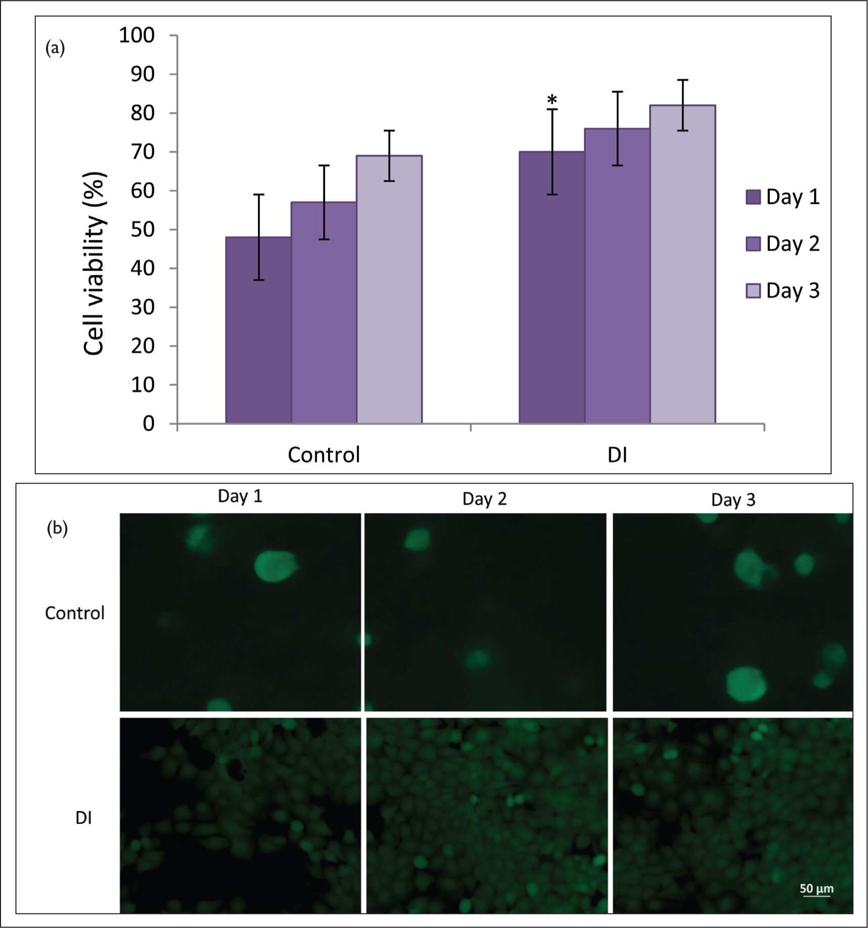
In this research, we introduce a DI that contains Si-NPs with the potential for bone healing. The prepared DI has shown outstanding mechanical properties, porous structure, bioactivity test, and biocompatibility study. Studies done in vitro showed that the implant increased cell viability, making it the best substrate for bone tissue engineering. Overall, the DI offers potential uses in bone tissue engineering, including the promotion of bioactive materials, dental soft and hard tissue healing, and new bone tissue formation. The present difficulty is scaling up the manufacturing of collagen and making it inexpensive for the general population while effectively incorporating MCF and Si-NPs into the development of nano-based biomaterials.

Ethical Approval

Not applicable

Informed Consent

Not applicable

Declaration of Conflicting Interests

The authors declared no potential conflicts of interest with respect to the research, authorship, and/or publication of this article.

Funding

The authors received no financial support for the research, authorship, and/or publication of this article.

References

1. Okuyama K, Miyama K, Mizuno K, et al. Crystal structure of (Gly-Pro-Hyp)9: Implications for the collagen molecular model. Biopolym. 2012;97:607–616.
2. Depboylu FN, Yasa E, Poyraz O, et al. Titanium based bone implants production using laser powder bed fusion technology. J Mater Res Technol. 2022;17:408–426.
3. Zafar MS, Khurshid Z, Almas K. Oral tissue engineering progress and challenges. Tissue Eng Regen Med. 2018;12:387–397.
4. Javed F, Akram Z, Khan J, et al. Growth factors and guided bone regeneration, Dental Implants, Woodhead Publishing Series in Biomaterials. 2020:133–143.
5. Janjua OS, Qureshi SM, Shaikh MS, et al. Autogenous Tooth Bone Grafts for Repair and Regeneration of Maxillofacial Defects: A Narrative Review. Int J Environ Res Public Health. 2022;19:3690.
6. Morgan EF, Unnikrisnan GU, Hussein AI. Bone mechanical properties in healthy and diseased states. Annu Rev Biomed Eng. 2018;20:119.
7. Omar F, Rozalia D, Charlotte AC, et al. The use of bone graft substitutes in large cancellous voids: Any specific needs?. Injury. 2011;42:87–90.
8. Yan C, Hao L, Hussein A, et al. Ti-6Al-4V triply periodic minimal surface structures for bone implants fabricated via selective laser melting. J Mech Behav Biomed Mater. 2015;51:61–73.
9. Pjetursson BE, Heimisdottir K. Dental implants–are they better than natural teeth? Eur J Oral Sci. 2018;126:81–87.
10. Senthil R, Sinem Ç, Batıkan Kavukcu S, et al. Fabrication of cylindrical bone graft substitute supported by reduced graphene-oxide and nanocurcumin to promotes the bone tissue development. Mater Lett. 2022;322:132475.
11. Gupta R, Gupta N, Weber, et al. Dental Implants. StatPearls Publishing; 2023.
12. Osuchukwu OA, Salihi A, Abdullahi I, et al. Synthesis techniques, characterization and mechanical properties of natural derived hydroxyapatite scaffolds for bone implants: a review. SN Appl Sci. 2021;3:822.
13. Senthil R, Kavukcu SB. Efficacy of glycoprotein-based nanocurcumin/silk fibroin electrospun scaffolds: Perspective for bone apatite formation. Mater Chem Phys. 2022;2899:126444.
14. Viguet-Carrin S, Garnero P, Delmas PD. The role of collagen in bone strength. Osteoporos Int. 2006;17:319–336.
15. Long FX. Building strong bones: Molecular regulation of the osteoblast lineage. Nat Rev Mol Cell Biol. 2012;13:27–38.
16. Ghosh S, Webster TJ. Mesoporous Silica Based Nanostructures for Bone Tissue Regeneration. Front Mater. 2021:8.
17. De Long WG, Einhorn TA, Koval K, et al. Bone, grafts and bone graft substitutes in orthopedic trauma surgery: A critical analysis. J Bone Joint Surg-Ame. 2007;89:0021–9355.
18. Du T, Niu Y, Liu Y, et al. Physical and Chemical Characterization of Biomineralized Collagen with Different Microstructures. J Funct Biomater. 2022;13:57.
19. Abd-elmohsen SA, Mohmed SA, Daigham GE, et al. Green synthesis, Optimization and Characterization of SiO2 nanoparticles using Aspergillus tubingensis F20 isolated from drinking water. Res Microbiol Journal. 2019;3:546–557.
20. Gour A, Jain NK, Advances in green synthesis of nanoparticles. Artificial cells. Nanomed Biotechnol. 2019;47:844–851.
21. Barzinjy A. Structure, synthesis and applications of ZnO nanoparticles: A review. Jordan J Phys. 2020;13:123–135.
22. Hodoroaba VD, Rades S, Salge T, et al. Characterisation of nanoparticles by means of high-resolution SEM/EDS in transmission mode. IOP Conf. Series: Mater Sci Eng 2016;109:012006.
23. Ye Y, Zhou Y, Sun Y, et al. Structure and infrared emissivity of collagen/SiO2 composite. Appl Surf Sci. 2008;254:5975–5980.
24. Senthil R, Basaran B, Vijayan S, et al. Electrospun nano-bio membrane for bone tissue engineering application: a new approach. Mater Chem Phys. 2020;249:123010.
25. Chen Y, Frith JE, Dehghan-Manshadi A, et al. Mechanical properties and biocompatibility of porous titanium scaffolds for bone tissue engineering. J Mech Behav Biomed Mater. 2017;75:169–174.
26. Velu R, Calais T, Jayakumar A, et al. A comprehensive review on bio-nanomaterials for medical implants and feasibility studies on fabrication of such implants by additive manufacturing technique. Materials. 2019;13:92.
27. Kokubo T, Takadama H. How useful is SBF in predicting in vivo bone bioactivity? Biomaterials. 2006;27:2907–2915.
28. Srinivasan S, Jayakumar R, Chennazhi K, et al. Multiscale Fibrous Scaffolds in Regenerative Medicine, Biomedical Applications of Polymeric Nanofibers. Springer; 2011:1–20.